
Animal Cell Diagram
An animal cell is a tiny, busy factory. This interactive diagram shows the main organelles — nucleus, mitochondria, endoplasmic reticulum, Golgi apparatus, lysosomes, ribosomes and more. Click any part to read what it does, hide the labels to test yourself, and print a labelled or blank copy for revision.
- Free to use
- No sign-up
- Runs in your browser
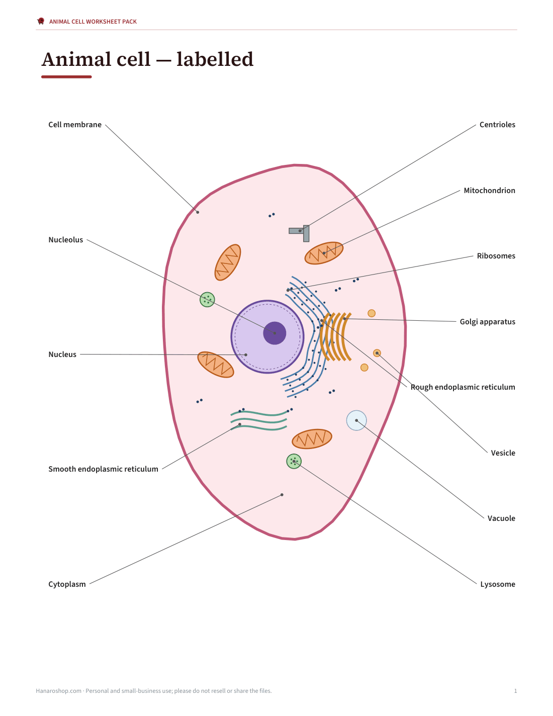
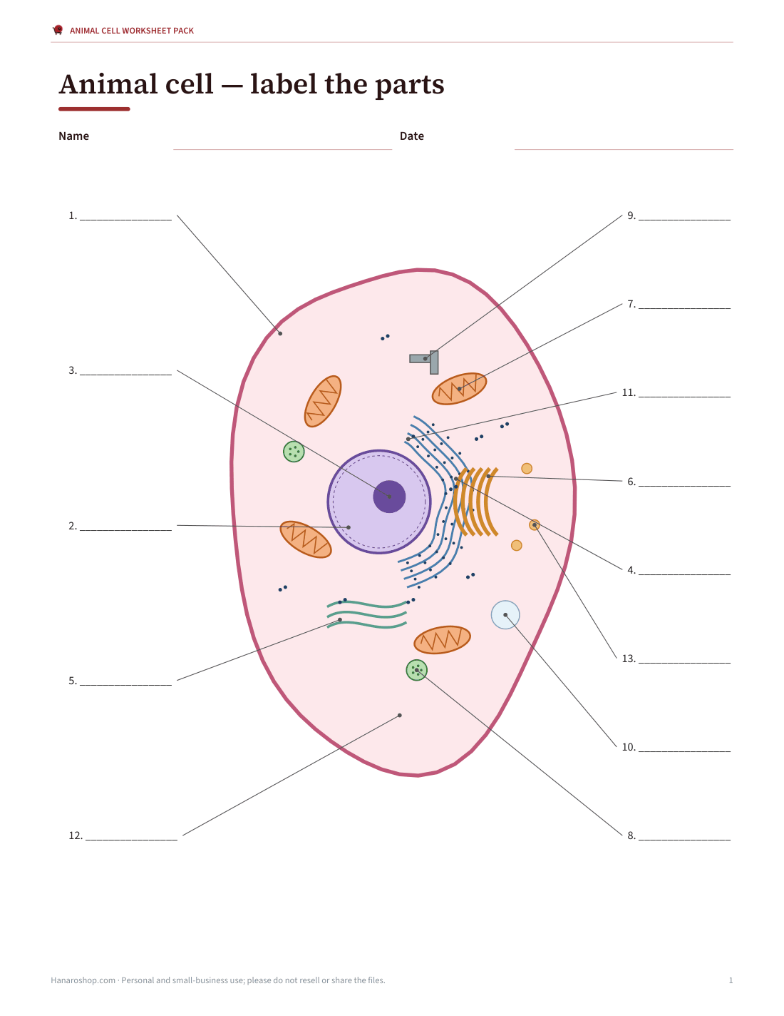
Interactive animal cell diagram
Animal Cell Worksheet Pack
Animal cell printables with answer keys: labelled diagram, blank numbered diagram, organelle function card sort, plant vs animal comparison, cloze worksheet and a 15-question quiz.
- Labelled diagram (PDF)
- Blank diagram (PDF)
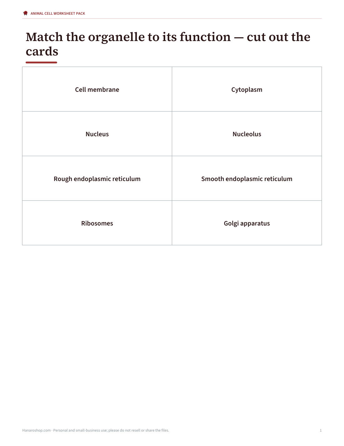
- Card sort (PDF)
- Functions table (PDF/DOCX)
- Plant vs animal (PDF, DOCX)
- Cloze worksheet (PDF, DOCX)
- 15-question quiz (PDF, DOCX)
Formats: PDF, DOCX. Instant download after payment (link valid 72 hours, up to 5 downloads). AI-assisted: the templates were drafted with AI help and reviewed and laid out before publishing.
$5.00 USD, one-time 7 files · about $0.71 each
- Instant download
- 7-day refund
- Secure checkout by Stripe
7-day money-back guarantee. Full refund within 7 days of purchase, even after you download.
Free sample: Animal Cell Labelled (PDF, 71 KB)
Full refund within 7 days — see the refund policy and license.
Free printables
Single sheets you can print right now, no sign-up. The full pack adds every sheet in one download plus the editable files.
 Printable Animal Cell Diagram Labelled Chart1 page · free PDF
 Printable Animal Cell Diagram2 pages · free PDF
 Animal Cell Organelle Card Sort2 pages · free PDF
 Printable Animal Cell Functions Chart1 page · free PDF
 Printable Plant vs Animal Cell Worksheet2 pages · free PDF
 Animal Cell Cloze Worksheet2 pages · free PDF
 Printable Animal Cell Quiz Sheet2 pages · free PDF
Parts of an animal cell and what they do
| Organelle | Function |
|---|---|
| Cell membrane | A thin, flexible phospholipid bilayer with embedded proteins that surrounds the cell and controls what enters and leaves. |
| Cytoplasm | The jelly-like fluid (cytosol) that fills the cell and holds the organelles; many chemical reactions happen here. |
| Nucleus | Contains the cell’s DNA and controls its activities; surrounded by a double nuclear envelope with pores. |
| Nucleolus | A dense region inside the nucleus that makes ribosomal RNA and assembles ribosome subunits. |
| Rough endoplasmic reticulum | Folded membranes studded with ribosomes that make and fold proteins, many of them for export or for membranes. |
| Smooth endoplasmic reticulum | Tube-like membranes without ribosomes that make lipids, help detoxify substances and store calcium. |
| Ribosomes | Tiny structures, free in the cytoplasm or on the rough ER, that build proteins by reading messenger RNA. |
| Golgi apparatus | A stack of flattened sacs that modifies, sorts and packages proteins and lipids into vesicles. |
| Mitochondrion | The site of aerobic respiration, releasing energy from glucose to make ATP; has its own DNA. |
| Lysosome | A small sac of digestive enzymes that breaks down waste, worn-out organelles and engulfed particles. |
| Centrioles | A pair of cylinders of microtubules near the nucleus that help organise the spindle during cell division. |
| Vesicle | A small membrane bubble that carries substances around the cell and to the membrane. |
| Vacuole | In animal cells, small and often temporary sacs for storage or transport (plant cells have one large vacuole). |
How the organelles work together
The organelles form a production line. Instructions stored in the DNA of the nucleus are copied into messenger RNA, which leaves through nuclear pores. Ribosomes — many of them on the rough endoplasmic reticulum — read the messages and build proteins. The proteins are folded in the ER, carried in vesicles to the Golgi apparatus, modified and sorted, then shipped in new vesicles to the cell membrane or to lysosomes. Mitochondria supply the energy (as ATP) for all this work, and lysosomes recycle worn-out parts.
Animal cells vs plant cells
| Feature | Animal cell | Plant cell |
|---|---|---|
| Cell wall | No | Yes (cellulose) |
| Chloroplasts | No | Yes |
| Vacuole | Small, several or none | One large central vacuole |
| Shape | Irregular, flexible | Fixed, often rectangular |
| Centrioles | Yes | Usually absent in flowering plants |
| Lysosomes | Common | Rare (vacuole does similar work) |
| Energy storage | Glycogen | Starch |
Both are eukaryotic cells with a nucleus, cell membrane, cytoplasm, mitochondria, ribosomes, endoplasmic reticulum and Golgi apparatus.
Using the interactive diagram
- Labelled mode shows every organelle with its name — click a part to read its function.
- Numbered mode hides the names so you can write them on a printout.
- Quiz mode describes a function and asks you to choose the organelle.
- Print any mode for revision notes or classwork.
Memory tips
- “Mighty mitochondria” — the powerhouse that makes ATP.
- Rough ER is rough because it is covered in ribosomes.
- Think of the Golgi as the post office: it packages and addresses parcels (vesicles).
- Lysosomes “lyse” (break down) — they are the recycling centre.
- The nucleolus lives inside the nucleus and makes ribosome parts.
Worked example
A Year 8 student revising for a biology test uses labelled mode to read about each organelle, then switches to quiz mode and scores 9 out of 12. They mix up smooth and rough ER, so they print the numbered diagram, label it from memory, and check against the labelled version — then retake the quiz and score 12.
What the printable pack contains
The template pack includes a labelled animal cell diagram, a blank numbered diagram for labelling, an organelle function card sort, a plant vs animal cell comparison worksheet, a cloze (fill-in-the-blanks) worksheet and a 15-question quiz, each with an answer key. It is designed for ages 11–16 and introductory biology courses.
How big is an animal cell?
Most animal cells are between about 10 and 30 micrometres across — roughly a tenth to a third of the thickness of a human hair — although there is huge variation: a human egg cell is about 0.1 mm and just visible to the naked eye, while some nerve cells have fibres over a metre long. The nucleus is typically 5–10 micrometres wide, mitochondria are 0.5–1 micrometre wide, and ribosomes are only about 20–30 nanometres, far too small to see with a light microscope.
A note on diagrams
Real cells are three-dimensional, crowded and vary widely by type — a nerve cell, a muscle cell and a white blood cell look very different. Textbook diagrams, including this one, are simplified models that show the typical organelles in one place and are not to scale. Under a light microscope you usually see only the membrane, cytoplasm and nucleus; most organelles need an electron microscope.
AI-assisted content
Text and worksheets were drafted with AI assistance and reviewed before publishing against standard school biology content. The diagram is a simplified illustration.
Frequently asked questions
What are the main parts of an animal cell?
Cell membrane, cytoplasm, nucleus, mitochondria, ribosomes, endoplasmic reticulum, Golgi apparatus, lysosomes and centrioles.
What is the powerhouse of the cell?
The mitochondrion, which releases energy as ATP through aerobic respiration.
Do animal cells have a cell wall?
No, only a flexible cell membrane.
What is the difference between rough and smooth ER?
Rough ER has ribosomes and makes proteins; smooth ER has none and makes lipids.
Can I print a blank animal cell diagram?
Yes, choose numbered mode and print.
What does the Golgi apparatus do?
It modifies, sorts and packages proteins and lipids into vesicles for transport.
Do all animal cells look the same?
No, cell shapes vary with their job, but most share the same set of organelles.
Is the diagram to scale?
No, it is a simplified model for learning.